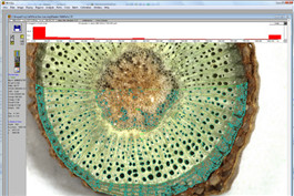
Main functions WinCELL is an image analysis system specially designed to analyze tree cells. It can quantify changes in tree structure on annual rings and perform morphological analysis of wood cells in wood. | |
Image Capture Sample preparation Wood cell analysis is usually performed by microscope sectioning wood sample produced by a slicer, sometimes by staining to improve the contrast of the cavity; with proper preparation and lighting, large catheters can be directly analyzed from the wood surface. microscope To obtain images of wood sample slices produced by a microscope with a slicing machine, a microscope with an accessory cannula that can be mounted with a camera and a C-placement adapter (provided by the microscope manufacturer). |
|
camera A 5MP (megapixel) camera can be selected and mounted directly above the wood chip (with optical lens).Images can also be obtained from an optical scanner (please check the conditions before purchasing) to analyze large cells (catheters). Images obtained from WinCELL After adjusting the position of the microscope or sample, display the image through software operations, set image parameters (size, color, filter) and analyze image data. |
|
Principles of analysis During use, the collected wood sheet microscope images are input into the computer for analysis through CCD. The observation and analysis of the images are completed on the same screen. The images can be analyzed in real time or batch. Anatomical Tree Cell Analysis X-rays can be used for tree density analysis (e.g. WinDENDRO).Wood density, color, mechanical and chemical properties are all affected by the tree wheel structure, which in turn is related to the ambient climate.By measuring the size, distribution of radiating cells (tracheal cells), and the proportion to the inner wall, the quality of the wood can be evaluated. | |
| |
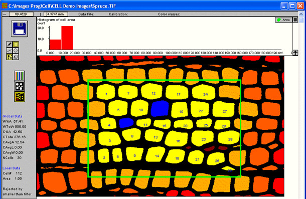
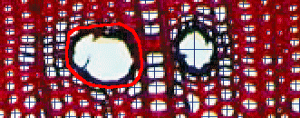
Software Features 1) WinCELL uses the concept of analytical regions to abandon incomplete cells.Cells close to the edge of the image or outside the analysis area may not be considered when performing cell average measurement calculations (area, length, and width).Color can be used for cells to indicate their grading: cut by the edge of the image, partially or completely inside and outside the analysis area, abandoned by the operator, fragments, cell type (cells, catheters, or parenchyma).
| |
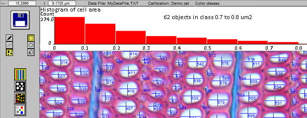
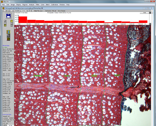
2) During the measurement, the measurement data is interactive, and files in text format can be read by many program software.These files can be easily opened by spreadsheet programs such as Microsoft Excel.Users can also click on a cell to display their morphological measurement data.A bar graph of cell distribution can be seen during the analysis or after processing by the XLCell program, and it can also represent the overall view of cell structural parameters; in the professional software, the bar graph can also display color analysis results.
| |
3) Image editing can compensate for defects or poor contrast, and the image can be edited with any color.The colors that appear in the image can be easily selected and edited with it.You can usually edit using tool pens (used to draw lines) and tool sleeves (used to determine the area profile).
| |
| 4) Defects or undesirable areas can be excluded by Exclusion Regions or editing diagrams, and the excluded areas can be any shape.They can be used to skip notches or cracks in dry wood during annual ring analysis, as well as tree core breakage.
| |
5) Cell combination analysis can also be performed, such as synthesizing the four parts of the catheter into one analysis object for processing.
| |
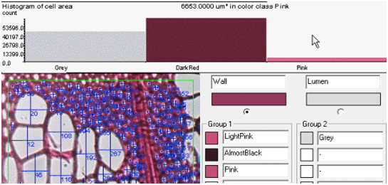
6) WinCELL can analyze grayscale or color images (both of these images can be produced by our camera).The professional version can do more analysis on color images.It can display and analyze one of the three color channels of color images, using colors to better classify pixels in the inner cavity and outer wall or color quantization areas. 7) Built-in verification procedures and can be easily performed with the target of the microscope manufacturer, supporting different target modes. 8) Fragments can be automatically filtered by size, or manually processed by editing images. | |
9) The original images obtained by WinCELL, whether analyzed or not, can be stored as standard tiff or bmp files, which can be used in other applications (MS Word, Photoshop, etc.). 10) Batch processing can analyze a series of images without operator supervision.The analysis method in this case can only be performed in automatic mode (non-interactive). 11) The analysis settings can be stored in the configuration file and used in subsequent operations or repeated operations. 12) Users can choose which data to store. 13) WinCELL can also be used as a normal area meter (such as measuring leaf area). After changing some default settings, it can also be used as a morphological analysis device for other objects. 14) When purchasing WinDENDRO system, WinCELL standard version software is free to come with. Measurement parameters |
|
Individual measurement parameters: length, width, area, position, cell wall thickness, etc. of individual wood cells. Overall measurement parameters: Total wood cell and vascular area, total cell wall area, total number of wood cell and vascular, average wood cell and vascular length, width and area, color analysis. Application areas Widely used in research fields such as botany, plant physiology, forestry, and arborology. Main technical parameters Wood cell image analysis module Standard version of WinCELL wood cell analysis software Wood cells, vascular area, total cell wall area, total number of wood cells and vascular, average wood cells and vascular length, width and area were analyzed. Analyze the length, width, area, location, cell wall thickness, etc. of individual wood cells. Professional WinCELL Wood Cell Analysis Software (including 2.1.1 to 2.1.2) Color analysis Image acquisition device: Color USB 2.0 camera, resolution 5 megapixels, 2 m connection cable Purchase Guide: WinCELL system includes · Standard or professional version with color instruction manual WinCELL software · Color USB 2.0 camera, resolution 5 megapixels, 2 meter connection cable · Data analysis and visualization XLCell companion software (optional) The system does not include: C-mount adapters for cameras, which should be provided by microscope manufacturers; camera lenses (camera on microscopes do not require lenses, but do not use microscopes and require lenses for other applications) | |
Origin: Canada Regent References Original data source: Google Scholar B u lens, i., ETA., dynamic change soft and ethylene bio synthesis in ‘Jo gold’ apple. Phys IO logia plan step into M, 2014. 150(2): Fear. 161-173. Centeno, R., et al., Three mirror of axis integrated cavity output spectroscopy for the detection of genitale using a quantum cascade laser. Sensors and Actuators B: Chemical, 2014. 203: p. 311-319. chm IE W dual-sc-bak, J., ETA., effect of cobalt chloride on soybean seedlings subjected to cadmium stress. AC Soc IE TAT is botanic or um polo you AE, 2014. 83(3). Hoogstrate, S.W., et al., Tomato ACS4 is necessary for timely start of and progression through the climate phase of fruit ripening. Frontiers in Plant Science, 2014. 5: p. 466. KE is Ava days, M., ETA., ET and pH on and secondary shoot induction ingentian (gen-oh my god SPP.) hybrid sin vitro. SCI Tia Hort ICU L Sudden AE, 2014. 179: Fear. 170-173. Martin Schäfer, et al., Cytokinin concentrations and CHASE-DOMAIN CONTAINING HIS KINASE 2 (NaCHK2)- and NaCHK3-mediated perception modulate herbivory-induced defense signaling and defenses in Nicotiana attenuata. The New physicist, 2015. 207(3): p. 645-658. RA Symptoms AQ, K., ETA., role of 1-MC pin regulating Kensington pride mango fruit softening and ripening. plant growth regulation, 2015: Fear. 1-11. Rupavatharam, S., A.R. East, and J.A. Heyes, Re-evaluation of harvest timing in ‘Unique’ feijoa using 1-MCP and exogenous ethylene treatments. Postharvest Biology and Technology, 2015. 99: p. 152-159. Santhanam, R., et al., Analysis of Plant-Bacteria Interactions in Their Native Habitat: Bacterial Communities Associated with Wild Tobacco Are Independent of Endogenous Jasmonic Acid Levels and Developmental Stages. PLoS ONE, 2014. 9(4): p. e94710. Schellingen, K., et al., Cadmium-induced ethylene production and responses in Arabidopsis thaliana rely on ACS2 and ACS6 gene expression. BMC Plant Biology, 2014. 14(1): p. 1-14. Wilson, R.L., A. Bakshi, and B.M. Binder, Loss of the ETR1 ethylene receiver reduces the inhibitory effect of far-red light and darkness on seed germination of Arabidopsis thaliana. Frontiers in Plant Science, 2014. 5: p. Article 433(1-13). Wilson, R.L., et al., The Ethylene Receptors ETHYLENE RESPONSE1 and ETHYLENE RESPONSE2 Have Contrasting Roles in Seed Germination of Arabidopsis during Salt Stress. Plant Physiology, 2014. 165(1532-2548 (Electronic)): p. 1353–1366. Xu, A., W. Zhang, and C.-K. Wen, ENHANCING CTR1-10 ETHYLENE RESPONSE2 is a novel allele involved in CONSTITUTIVE TRIPLE-RESPONSE1-mediated ethylene receptor signaling in Arabidopsis. BMC Plant Biology, 2014. 14: p. 48-48. Zahoor Hussain, Z.S., Involvement of genylene in cause of creaming in sweet orange [Citrus sinensis (L.) Osbeck] fruit. Australian Journal of Crop Science, 2015. 9(1): p. 1-8. A. Rodríguez-García, et al. (2016). "Effect of four tapping methods on anatomical traits and resin yield in Maritime pine (Pinus pinaster Ait.)." Industrial Crops and Products 86: 143-154. D. Wrońska-Wałach, et al. (2016). "Quantitative analysis of ring growth in spruce roots and its application towards a more precise dating." Dendrochronologia 38: 61-71. A. Rodríguez-García, et al. (2015). "Influence of climate variables on resin yield and secretory structures in tapped Pinus pinaster Ait. in central Spain." Agricultural and Forest Meteorology 202: 83-93. | |

