1. Product functions
MoniCyte live cell intelligent monitoring system is small in size and can be placed in the incubator to monitor live cell growth. The system is equipped with powerful AI analysis function to quickly synthesize dynamic videos of cell growth, analyze cell density, convergence, and draw cell growth curves. No one is present.Monitor cell growth on duty.It can be applied in the fields of cell biology such as tumor cell migration, nerve cell growth, stem cell differentiation, cytotoxicity experiments, egg cell maturation, organoid growth, etc.
2. Product Features
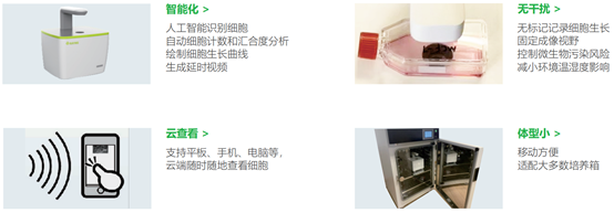
Intelligence: Artificial intelligence recognizes cells, automatically analyzes cell counting and confluence, draws cell growth curves, and generates time-lapse videos
No interference: record cell growth without labels, fix imaging field, control the risk of microbial contamination, reduce the impact of environmental temperature and humidity
Meta view: Supports tablets, mobile phones, computers, etc., and view cells anytime, anywhere in the cloud
Small size: easy to move, suitable for most incubators

III. Application direction
Tumor immunity: 1. New drug development/drug screening-Medical chemistry and toxicology research: cytotoxicity experiments of drugs (Traditional medicine), evaluation of tumor cell migration ability, scratch experiments.2. Cell therapy: immune cell expansion, immune killing experiments, phagocytosis.
Cell culture: 1. Cell culture, stem cell proliferation and differentiation process: Cell culture QC, draw cell growth curve, and synthesize time-lapse video.2. Cell construction: cell transfection efficiency, reporter gene cell line construction.
Organoids: Monitor tumor sphere formation, growth and health status in real time.
Reproductive genetics: egg cell/embryo differentiation.
4. Model parameters
MC-B100: Bright-field imaging, suitable for most bright-field label-free cell imaging scenarios
1. Bright field lighting: LED
2. Fluorescence channel:/
3. Magnification: 10× fixed objective lens
4. Imaging field of vision: single field of vision
5. Camera: 6MP CMOS
6. Objective lens focus: electric
7. Culture container: multi-well plates, petri dishes, culture bottles, slides and other transparent containers with a height of less than 50 mm
8. Size: 150×170×180 mm (L×W×H)
9. Weight: 2kg
10. Working environment: Temperature: 5℃-40℃; Humidity: 20%-95%
MC-F100: Standard red and green fluorescence channels to meet fluorescence imaging application scenarios such as cell transfection and cell line construction
1. Bright field lighting: LED
2. Fluorescence channel: dual-channel fluorescence.Red: Ex=530/15nm, Green: Ex=470/10nm
3. Magnification: 10× fixed objective lens
4. Imaging field of vision: single field of vision
5. Camera: 5MP CMOS
6. Objective lens focus: electric
7. Culture container: multi-well plates, petri dishes, culture bottles, slides and other transparent containers with a height of less than 50 mm
8. Size: 220×200×220 mm (L×W×H)
9. Weight: 4kg
10. Working environment: Temperature: 5℃-40℃; Humidity: 20%-95%
MC-S100: High-throughput bright-field imaging, single-well or whole plate scanning, long-term monitoring of live cells without labeling.
1. Bright field lighting: LED
2. Fluorescence channel:/
3. Magnification: 10× fixed objective lens
4. Imaging field of view: Whole plate scanning (86mm×127mm)
5. Camera: 5MP CMOS
6. Objective lens focus: electric
7. Culture container: multi-well plates, petri dishes, culture bottles, slides and other transparent containers with a height of less than 50 mm
8. Size: 368×350×242 mm (L×W×H)
9. Weight: 9kg
10. Working environment: Temperature: 5℃-40℃; Humidity: 20%-95%
Among them, MC-B100 and MC-F100 support remote access and analysis functions in the cloud.
Manufacturer: Ruiming Bio