Product Features
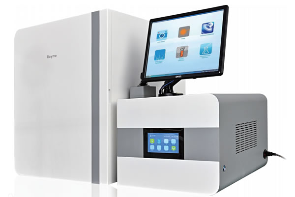
The real-time single-cell multifunctional analysis system can detect the metabolic small molecule content and enzyme activity of a single living cell in real time, continuously and quantitatively.This product complements the need for new detection technologies for single-cell research, helping users better understand the diversity of cell composition, physiological behavior and functions.

Main performance
● Real-time single-cell multi-indicator detection: Real-time detection of small molecules in a single living cell (such as glucose, lactic acid, ATP, cholesterol, C+, K, etc.) and enzyme activities (glucosidase, sphingomyelinase, lactate dehydrogenase, etc.), can match more than 100 kinds of commercial kits;
● Real-time subcellular in situ detection: Real-time continuous and in situ detection at the subcellular level (cytoplasm, nucleus, and membrane);
● Ultramicro extraction and injection: Extract organelles (such as lysosomes, mitochondria) and cytoplasm at a single cell level for mass spectrometry or other platforms for combined use analysis; inject drugs, metabolic agents, etc. in a single cell, and perform drug efficacy evaluation;
● Live level detection: Real-time detection of changes in biochemical indicators (before and after medication, before and after stimulation of traditional Chinese medicine acupuncture).
Features
● Real-time detection of small molecules and enzyme activity in a single living cell can be carried out;

Controllable light activation enables ATP detection for single living cells with high spatiotemporal resolution
zh Volume D uPressETA..analytical chemistry. 2021.

Analysis of enzyme activity in single cells to improve understanding of cell heterogeneity and signaling cascade
RP PressETA..P NAS.2016.
● The extraction and injection module can realize single-cell site extraction and drug injection analysis;

Aldometanib ("fasting") activated AMPK through "lysosomal pathway" and experimental results of real-time single-cell multifunctional analysis system: Ultramicromicro injection of FBP (1,6-phosphate fructose) failed to restore AldometanibThe fluorescence signal of TRPV4-GCaMP6s in pretreated cells, while FBP restored the fluorescence signal in control cells that were not treated with fasting, proving that fasting can effectively inhibit the binding of FBP to aldolase.
Zhang CS, l IM, Wang Y,ETA..nature metabolism. 2022.

Single lysosome extraction at subcellular level and achieve glucosidase activity analysis
RP PressETA..Proc. Natl. Acad.SCI.2018.

Subcellular level and targeted drug delivery and real-time monitoring and analysis
TZ X inETA..S code.2012.
●The dual function matching of fluorescence detection and electrochemical detection can be achieved.

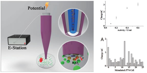
Fluorescence detection: The excitation light source stimulates the fluorescent substances of cells through the light probe (generated by fluorescence staining or other methods) to cause the cells to produce fluorescence; the optical detection system collects fluorescence signals, and the signal strength reflects the content of small molecules or enzyme activities in the cell.Electrical testing: The electrical probe directly detects the current value of the electroactive substances released in cells or cells, and the current intensity reflects the detection of small molecule content or enzyme activity.
Application direction
●Tumor mechanism: lung cancer, breast cancer, liver cancer
●New drug research: pharmacology, drug astronomy
●Neural mechanisms: Alzheimer's disease, Parkinson's disease, Huntington's disease
● Immune regulation: inflammation, anti-tumor
●Cardiovascular diseases: atherosclerosis, hypertension, hyperlipidemia
● Cell senescence: cell autophagy, mitochondria
Model parameters
●Ray-mini: Inverted bright field imaging.Color HD CMOS camera.Standard 4×, 10×, 20×, 40× objective lenses, manually switched.The minimum current is 10nA, and the current resolution is <1pA.The microscopic operating system has manual X, Y and Z axes, with a step of 13mm and a precision of 10μm; T-axis (oblique): manual, stroke of 100mm; R-axis (angle): manual, angle of 20°-90°, accuracy of 10’.Manual X-axis and Y-axis adjustment sample platform; adjustment accuracy: 10μm; stroke: 20mm.Software: Bright-field imaging, electrical signal acquisition and analysis.Optical, electrical and magnetic shielding.Appearance dimensions: all-in-one structure, 650×850×1000cm.The rated voltage is 220V, the power is <600W, and the weight is about 65kg.
●Ray-E00: Inverted fluorescence imaging.Color highly sensitive CMOS camera.Standard 4×, 10×, 20×, 40× objective lenses, manually switched.The minimum current is 10nA, and the current resolution is <1pA.The microscopic operating system is automatic with a step of 20mm and a precision of 50μm; T-axis (oblique): manual, stroke of 100mm; R-axis (angle): manual, angle of 20°-90°, accuracy of 10’.Manual X-axis and Y-axis adjustment sample platform; adjustment accuracy: 10μm; stroke: 20mm.Software: fluorescence imaging, microscopic operation control, electrical signal acquisition and analysis.Optical, electrical and magnetic shielding.Appearance dimensions: split structure: operating room 680×810×850mm; electrical room 500×660×850mm.The rated voltage is 220V, the power is <600W, and the weight is about 90kg.
●Ray-l00: Inverted fluorescence imaging.Color HD CCD camera.Standard 4×, 10×, 20×, 40× objective lenses, manually switched.Single channel (choose one of three channels, ultraviolet light, blue light, and green light), with a detection wavelength of 230-700nm.The sensitivity of the optical signal is ≥50 photons.The microscope operating system is manual, with a step of 13mm and a precision of 10μm; T-axis (oblique): manual, stroke of 100mm; R-axis (angle): manual, angle of 20°-90°, accuracy of 10’.Manual X-axis and Y-axis adjustment sample platform; adjustment accuracy: 10μm; stroke: 20mm.Software: Fluorescence imaging, optical signal acquisition and analysis.Optical, electrical and magnetic shielding.Appearance dimensions: split structure: operating room 680×810×850mm; electrical room 500×660×850mm.The rated voltage is 220V, the power is <600W, and the weight is about 90kg.
●Ray -M00: Inverted fluorescence imaging.Color HD CCD camera.Standard 4×, 10×, 20×, 40× objective lenses, manually switched.Three channels (optional with other light sources, ultraviolet, blue light, and green light), detection wavelength is 230-700nm.The sensitivity of the optical signal is ≥50 photons.The minimum current is 1pA, the current resolution is <0.01pA.The microscope operating system has the right and left hand positions, X, Y, Z axis manual and X, Y, Z axis invisible speed change, X, Y and Z axis manual: 13mm step, 10μm accuracy; X, Y and Z axis automatic: 20mm step, Accuracy 50nm; T-axis (oblique): manual, stroke 100mm; R-axis (angle): manual, angle 20°-90°, accuracy 10'.Manual X-axis and Y-axis adjustment sample platform; adjustment accuracy: 10μm; stroke: 20mm.Software: Fluorescence imaging, microscopic operation control: optical and electrical signal acquisition and analysis.Optical, electrical and magnetic shielding.Appearance dimensions: split structure: operating room 680×810×850mm; electrical room 500×660×850mm.The rated voltage is 220V, the power is <600W, and the weight is about 90kg.
●Ray -prod: Inverted fluorescence imaging.Monochrome high sensitivity CCD camera.Standard 4×, 10×, 20×, 40× objective lenses, manually switched.Three channels (optional with other light sources, ultraviolet, blue light, and green light), detection wavelength is 230-800nm.The sensitivity of the optical signal is ≥50 photons.The minimum current is 1pA, the current resolution is <0.01pA.It has ultra-micro extraction and injection function.The microscope operating system has fully automatic speed change of the X, Y, and Z axis of the left and right hand positions.X, Y and Z axes automatic: 20mm step, 50nm accuracy; T-axis (oblique): manual, stroke 100mm; R-axis (angle): manual, angle 20°-90°, accuracy 10’.Manual X-axis and Y-axis adjustment sample platform; adjustment accuracy: 10μm; stroke: 20mm.Software: fluorescence imaging, microscopic operation control, optical and electrical signal acquisition and analysis, ultramicro extraction and injection monitoring.Optical, electrical and magnetic shielding.Appearance dimensions: split structure: operating room 680×810×850mm; electrical room 500×660×850mm.The rated voltage is 220V, the power is <600W, and the weight is about 90kg.
●Ray-high D: Inverted fluorescence imaging.Monochrome high sensitivity CCD camera.Standard 4×, 10×, 20×, 40×, 60× objective lenses are equipped with automatic switching.Three channels (optional with other light sources, ultraviolet, blue light, and green light), detection wavelength is 230-800nm.The sensitivity of the optical signal is ≥50 photons.The minimum current is 1pA, the current resolution is <0.01pA.It has ultra-micro extraction and injection function.The microscope operating system has fully automatic speed change of the X, Y, and Z axis of the left and right hand positions.X, Y and Z axes automatic: 20mm step, 50nm accuracy; T-axis (oblique): manual, stroke 50mm; R-axis (angle): manual, angle 20°-90°, accuracy 10’.Fully automatic adjustment of X-axis and Y-axis electric stages, stroke: 100×50mm, displacement resolution 0.1μm.Software: fluorescence imaging, microscopic operation control, optical and electrical signal acquisition and analysis, ultramicro extraction and injection monitoring.Fully automatic intelligent cell detection.Optical, electrical and magnetic shielding.Appearance dimension: Integral structure: 1000×530×849mm. Rated voltage is 220V, power is <600W, weight is about 120kg.
factory: Ruiming Bio